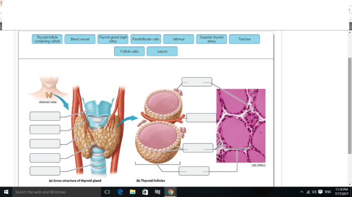
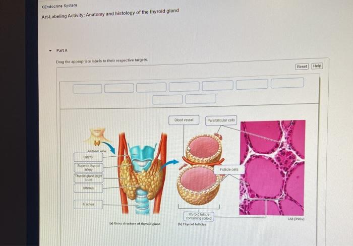
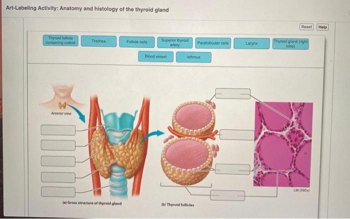
Art labeling activity anatomy and histology of the thyroid gland – Embark on an immersive journey with our art labeling activity that unveils the intricacies of the thyroid gland’s anatomy and histology. Prepare to delve into a realm of scientific exploration, where art and knowledge intertwine, fostering a profound understanding of this essential endocrine organ.
The thyroid gland, nestled within the anterior neck region, plays a pivotal role in regulating metabolism, growth, and development. Its unique structure and cellular composition, meticulously detailed in this activity, will captivate your curiosity and enhance your comprehension of its vital functions.
Anatomy of the Thyroid Gland: Art Labeling Activity Anatomy And Histology Of The Thyroid Gland

The thyroid gland is a small, butterfly-shaped endocrine gland located in the neck, just below the Adam’s apple. It consists of two lobes, one on each side of the trachea, connected by a narrow isthmus. The thyroid gland is surrounded by the strap muscles of the neck and is in close proximity to the parathyroid glands, esophagus, and recurrent laryngeal nerves.
Lobes and Isthmus
The thyroid gland is divided into two lateral lobes, which are connected by a thin, central isthmus. The lobes are roughly pyramidal in shape, with their bases facing the trachea and their apices extending laterally. The isthmus is located at the level of the second and third tracheal rings and connects the two lobes anteriorly.
Relationship to Surrounding Structures, Art labeling activity anatomy and histology of the thyroid gland
The thyroid gland is closely related to several important structures in the neck. The trachea, which carries air to and from the lungs, lies directly anterior to the thyroid gland. The esophagus, which carries food from the pharynx to the stomach, is located posterior to the thyroid gland.
The parathyroid glands, which regulate calcium metabolism, are located on the posterior surface of the thyroid gland. The recurrent laryngeal nerves, which innervate the muscles of the larynx, run along the posterior and lateral borders of the thyroid gland.
Histology of the Thyroid Gland

The thyroid gland is composed of follicles, which are spherical structures lined by a single layer of follicular cells. Follicular cells secrete thyroid hormones, which regulate metabolism. Parafollicular cells, also known as C cells, are located between the follicles and secrete calcitonin, which regulates calcium metabolism.
Follicles
Thyroid follicles are the functional units of the thyroid gland. They are lined by a single layer of follicular cells, which are cuboidal or columnar in shape. The follicles are filled with a viscous fluid called colloid, which contains the thyroid hormones thyroxine (T4) and triiodothyronine (T3).
Follicular Cells
Follicular cells are the main cells of the thyroid gland. They synthesize and secrete thyroid hormones, which regulate metabolism. Thyroid hormones are essential for growth, development, and reproduction.
Parafollicular Cells
Parafollicular cells, also known as C cells, are located between the follicles. They secrete calcitonin, which regulates calcium metabolism. Calcitonin lowers blood calcium levels by inhibiting osteoclasts, which are cells that break down bone.
Art Labeling Activity

Materials
* Image of the thyroid gland
- Markers or colored pencils
- Ruler or protractor
- Scissors
- Glue
Instructions
Print out an image of the thyroid gland.
2. Label the following structures on the image
Thyroid lobes
Isthmus
Trachea
Esophagus
Parathyroid glands
- Recurrent laryngeal nerves
- Color the thyroid gland and the surrounding structures.
- Cut out the image of the thyroid gland and glue it to a piece of paper.
- Write a short paragraph describing the anatomy of the thyroid gland.
FAQs
What is the significance of the thyroid gland?
The thyroid gland secretes hormones essential for regulating metabolism, growth, and development.
How does the art labeling activity enhance learning?
By combining visual art with anatomical labeling, the activity engages multiple learning pathways, promoting deeper comprehension and retention.
What are the different cell types found in the thyroid gland?
Follicular cells, parafollicular cells, and C cells, each with distinct functions in hormone production and regulation.