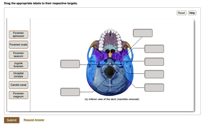
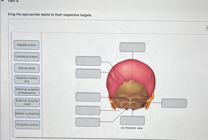
Drag the appropriate labels to their respective targets. skull – Embark on a captivating exploration of the human skull with our comprehensive guide, “Drag the Appropriate Labels to Their Respective Targets: Skull.” This interactive resource provides an immersive learning experience, empowering you to master the intricacies of skull anatomy, musculature, foramina, canals, imaging, and fractures.
Our meticulously crafted guide delves into the diverse components that constitute the skull, unraveling their functions and clinical significance. Engage with interactive diagrams, detailed descriptions, and thought-provoking discussions to gain an unparalleled understanding of this essential anatomical structure.
Skull Anatomy: Drag The Appropriate Labels To Their Respective Targets. Skull

The skull is a complex and fascinating structure that forms the framework of the head. It is made up of 22 bones that are joined together by sutures. The skull protects the brain, eyes, ears, and other important structures of the head.
It also provides attachment points for the muscles of the face and neck.
The skull is divided into two main parts: the cranium and the facial skeleton. The cranium is the upper part of the skull and it houses the brain. The facial skeleton is the lower part of the skull and it forms the face.
The cranium is made up of eight bones: the frontal bone, the parietal bones (2), the occipital bone, the temporal bones (2), the sphenoid bone, and the ethmoid bone. The facial skeleton is made up of 14 bones: the nasal bones (2), the maxillae (2), the zygomatic bones (2), the lacrimal bones (2), the palatine bones (2), the inferior nasal conchae (2), and the mandible.
The skull bones are joined together by sutures. Sutures are immovable joints that allow for some growth of the skull. The sutures are named after the bones that they connect. For example, the coronal suture is the suture that connects the frontal bone to the parietal bones.
The skull bones have a variety of functions. The cranium protects the brain from injury. The facial skeleton supports the muscles of the face and neck. The skull also provides attachment points for the ligaments and tendons of the head and neck.
Skull Musculature, Drag the appropriate labels to their respective targets. skull
The skull is attached to the body by muscles. These muscles are responsible for moving the head and neck. The muscles that attach to the skull are divided into two groups: the extrinsic muscles and the intrinsic muscles. The extrinsic muscles are the muscles that attach to the skull from outside the skull.
The intrinsic muscles are the muscles that attach to the skull from inside the skull.
The extrinsic muscles of the skull are the sternocleidomastoid muscle, the trapezius muscle, the splenius capitis muscle, the longus capitis muscle, and the rectus capitis anterior muscle. The sternocleidomastoid muscle is the large muscle that runs from the sternum to the mastoid process of the temporal bone.
The trapezius muscle is the large muscle that runs from the occipital bone to the clavicle and scapula. The splenius capitis muscle is the muscle that runs from the occipital bone to the cervical vertebrae. The longus capitis muscle is the muscle that runs from the cervical vertebrae to the occipital bone.
The rectus capitis anterior muscle is the muscle that runs from the atlas vertebra to the occipital bone.
The intrinsic muscles of the skull are the temporalis muscle, the masseter muscle, the pterygoid muscles, and the digastric muscle. The temporalis muscle is the large muscle that runs from the temporal bone to the mandible. The masseter muscle is the large muscle that runs from the maxilla to the mandible.
The pterygoid muscles are the muscles that run from the sphenoid bone to the mandible. The digastric muscle is the muscle that runs from the mandible to the hyoid bone.
The skull muscles work together to move the head and neck. The extrinsic muscles are responsible for moving the head in relation to the body. The intrinsic muscles are responsible for moving the mandible and the other bones of the skull.
Skull Foramina and Canals
The skull has a number of foramina and canals that allow for the passage of nerves and blood vessels. The foramina are openings in the skull bones. The canals are tunnels that run through the skull bones.
The major foramina of the skull are the foramen magnum, the foramen ovale, the foramen rotundum, the foramen spinosum, and the jugular foramen. The foramen magnum is the large opening in the occipital bone that allows for the passage of the spinal cord.
The foramen ovale is the opening in the sphenoid bone that allows for the passage of the mandibular nerve. The foramen rotundum is the opening in the sphenoid bone that allows for the passage of the maxillary nerve. The foramen spinosum is the opening in the sphenoid bone that allows for the passage of the middle meningeal artery.
The jugular foramen is the opening in the occipital bone that allows for the passage of the jugular vein and the glossopharyngeal, vagus, and accessory nerves.
The major canals of the skull are the optic canal, the internal carotid canal, the hypoglossal canal, and the facial canal. The optic canal is the canal in the sphenoid bone that allows for the passage of the optic nerve.
The internal carotid canal is the canal in the temporal bone that allows for the passage of the internal carotid artery. The hypoglossal canal is the canal in the occipital bone that allows for the passage of the hypoglossal nerve.
The facial canal is the canal in the temporal bone that allows for the passage of the facial nerve.
The skull foramina and canals are important for the passage of nerves and blood vessels. They allow for the brain to communicate with the rest of the body and for the skull to receive blood and nutrients.
Skull Imaging
There are a number of different imaging techniques that can be used to visualize the skull. These techniques include X-rays, computed tomography (CT) scans, and magnetic resonance imaging (MRI) scans.
X-rays are a type of radiation that can be used to create images of the skull. X-rays are often used to diagnose skull fractures and other injuries. CT scans are a type of X-ray that uses a computer to create cross-sectional images of the skull.
CT scans can be used to diagnose skull tumors and other abnormalities.
MRI scans are a type of imaging technique that uses magnets and radio waves to create images of the skull. MRI scans can be used to diagnose skull tumors, skull fractures, and other abnormalities. MRI scans are also used to study the brain and other structures of the head.
The different imaging techniques used to visualize the skull have different advantages and disadvantages. X-rays are a quick and inexpensive way to image the skull. However, X-rays can only show the bones of the skull. CT scans can provide more detailed images of the skull than X-rays.
However, CT scans are more expensive than X-rays and they can expose the patient to more radiation. MRI scans can provide the most detailed images of the skull. However, MRI scans are the most expensive and time-consuming of the imaging techniques.
Skull Fractures
Skull fractures are a break in one or more of the bones of the skull. Skull fractures can be caused by a variety of injuries, such as falls, blows to the head, and motor vehicle accidents.
There are three main types of skull fractures: linear fractures, depressed fractures, and compound fractures. Linear fractures are the most common type of skull fracture. They are caused by a blow to the head that causes the bone to crack.
Depressed fractures are caused by a blow to the head that causes the bone to be pushed inward. Compound fractures are caused by a blow to the head that causes the bone to break and the skin to be torn.
The signs and symptoms of a skull fracture can vary depending on the type of fracture. Linear fractures often cause only mild symptoms, such as a headache and bruising. Depressed fractures can cause more severe symptoms, such as nausea, vomiting, and seizures.
Compound fractures can cause the most severe symptoms, such as coma and death.
The treatment for a skull fracture depends on the type of fracture. Linear fractures often do not require treatment. Depressed fractures may require surgery to lift the bone back into place. Compound fractures may require surgery to repair the bone and the skin.
Frequently Asked Questions
What are the major bones that make up the skull?
The major bones of the skull include the frontal bone, parietal bones, temporal bones, occipital bone, sphenoid bone, ethmoid bone, nasal bones, maxillae, and mandibles.
How do the skull muscles contribute to facial expressions?
The muscles that attach to the skull play a crucial role in facial expressions by controlling the movement of the eyebrows, eyelids, nose, and mouth.
What is the clinical significance of the skull foramina and canals?
The skull foramina and canals provide passage for nerves and blood vessels, and their clinical significance lies in their role in diagnosing and treating various medical conditions.

